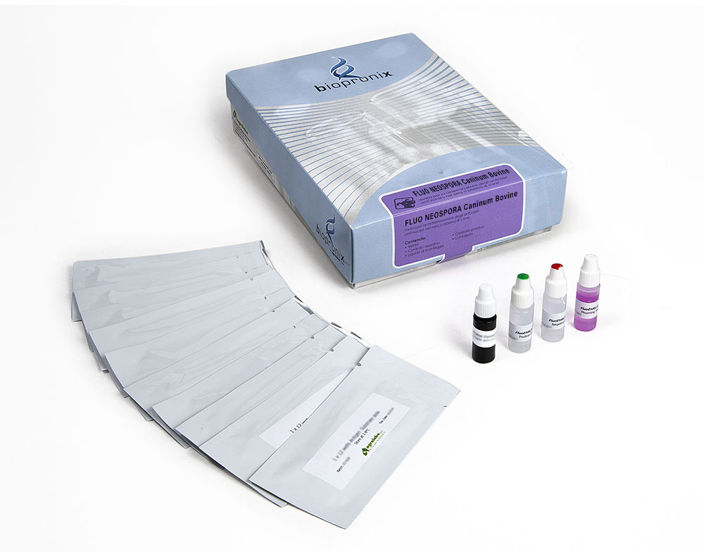
Fluo Neospora caninum bovine
IFA kit for the detection of anti-Neospora caninum IgG antibodies in bovine serum or plasma
50 slides
complete Kit
The FLUO NEOSPORA caninum BOVINE kit is intended for the semiquantitative detection of IgG antibodies to Neospora caninum in bovine serum or plasma by indirect immunofluorescence assay
Neospora caninum is a protozoan which is believed to be an important abortion agent in bovine species throughout the world. It is believed that it may represent, in some areas (eg Northern Australia), 30% of all causes of bovine abortion; the infection could affect over 20% of farms.
The economic damage is essentially linked to abortion (loss of the calf, lack of milk production, etc.), but a 5% decrease in milk production during the first lactation was also observed in infected cows that did not abort.
In addition to cattle, the existence of Neospora caninum infections has been identified in numerous other animal species, such as: dog, sheep, goat, horse, deer.
The main manifestation of the infection is represented by abortion, which typically occurs between the fourth and seventh months of gestation.
Occasionally, in calves, incoordination of movements and paralysis of the limbs may occur at birth, while in adults the infection is asymptomatic.
The frequency of abortions in the herd is highly variable. It is generally believed that abortion occurs occasionally (and therefore with low or very low frequency). In many farms, the infection goes unnoticed. However, episodes characterized by a very high incidence of abortions and in a relatively short period of time are also reported. In the most serious cases, abortion affected 1/3 of pregnant cows within a few months. It is hypothesized that these "waves" of abortions ("abortion storms"), which can also appear in herds where no reproductive problems had previously been reported, are triggered by immunosuppressive factors (eg presence of mycotoxins in the ration).
Infected cow does not always abort but often gives birth to a calf that is born already infected by transplacental infection. The infection is persistent (the infected animal never "heals", in the sense that the parasite persists in the body indefinitely), and therefore is transmitted from generation to generation.
Bovine sera are diluted in buffered saline and incubated in the individual slide wells to allow reaction of patient antibody with Neospora antigens on the wells.
Slides are then washed to remove unreacted serum proteins, a fluorescence-labelled FITC anti-bovine IgG (conjugate) is added. This conjugate is allowed time to react with antigen-antibody complexes. The slides are washed again to remove unreacted conjugate. The resulting reactions can be visualized using standard fluorescence microscopy, where a positive reaction is seen as sharply defined apple-green fluorescence of the whole membrane of Neospora tachyzoites. A negative reaction is seen either as a red fluorescence of the Neospora tachyzoites, or will show a pole fluorescence of the membrane of tachyzoites or fluorescence unlike that seen in the positive control well.
Positive reactions may then be retested at a higher dilution to determine the highest reactive (endpoint titer).
Bovine serum or plasma samples.
After collection, once coagulation has taken place, the serum is separated by centrifugation, then transferred (serum or plasma) into sterile tubes. Store at + 2-8 °C. If testing is performed after more than 5 days, samples should be frozen at -20 °C or colder. Samples taken from subjects with acute form must be collected at the beginning of the disease; further samples may be taken at intervals of two and four weeks to detect any change in antibody titer during convalescence.
To read the results, use the fluorescence microscope with FITC filter at 400X magnification.
The fluorescence pattern (shape, density, etc.) of the negative and positive control should be considered the reference model.
Reactivity patterns other than those present in the controls should be considered non-specific reactions (negative result).
The presence of anti-Neospora caninum antibodies depends on the geographic regions and populations analysed.
