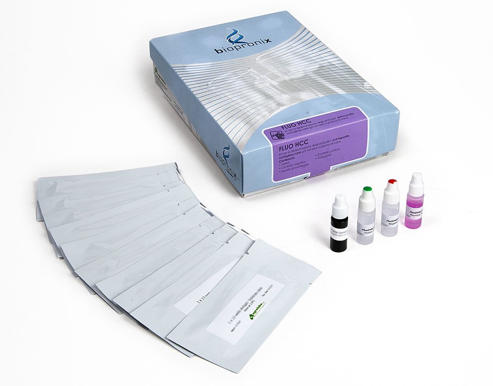
FLUO HEPATITIS contagiosa canis
IFA kit for the detection of IgG antibodies to infectious hepatitis virus
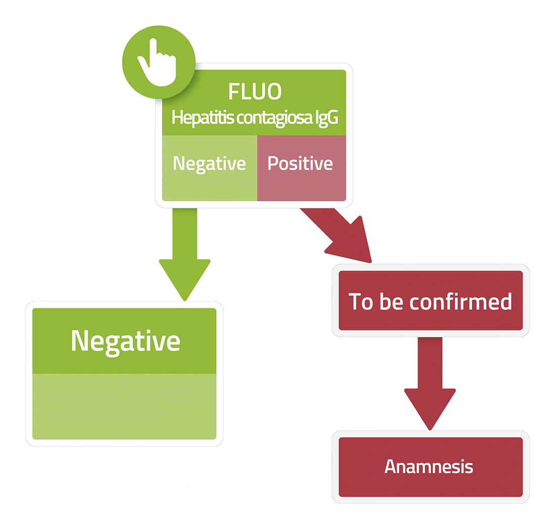
Fluo HEPATITIS contagiosa canis is a test based on the immunofluorescence technique for the detection of IgG antibodies to infectious hepatitis virus in dog serum or plasma samples.
Canine infectious hepatitis (formerly called Rubarth’s disease) is an acute liver infection caused by canine adenovirus type 1 (CAV-1). In Europe it mainly affects dogs and foxes.
It is a disease unrelated to hepatitis in humans, it is a much less common condition thanks to the availability of effective vaccines. However, from time to time this highly contagious and sometimes fatal disease is still observed in veterinary practice, particularly in unvaccinated puppies.
The main source of infection is the ingestion of urine, feces or saliva from infected dogs. After healing, dogs can spread the virus through their urine for up to 6 months. The virus has a predilection for hepatocytes, the vascular epithelium and the renal epithelium. Viral replication occurs in the tonsils and then spreads to the local lymph nodes and, via the systemic circulation, to the hepatic and endothelial cells.
Adenoviruses cause necrosis of infected cells by a cytopathic effect: lesions include petechiae, diffuse bruising, fibrin on the liver capsule, and possiblecollection of clear peritoneal exudate.
The virus is resistant to numerous disinfectants and can remain in the environment for several weeks or months.
Very young puppies die within hours, and in settings like dog shelters the disease spreads very quickly.
Symptoms can vary greatly, from mild to death, and different forms of the disease are observed.
Hyperacute form (in young puppies)
Puppies under 3 weeks of age may show a sudden swelling of the abdomen and succumb within a few hours. Most puppies from reputable sources have temporary immune protection inherited from their mother.
Acute form (classical disease)
In the initial stages of the disease, the dog is not very vital, with fever and inflammation of the tonsils (tonsillitis), as well as a very strong redness of the mucous membranes and swelling of the lymph nodes under the jaw.
The disease progresses rapidly to the onset of vomiting and/or diarrhea, with loss of appetite.
The liver is painful and enlarged on palpation. With the development of liver failure, jaundice and bleeding of the gums appear. At this stage of the disease, the membranes become pale or jaundiced. The dog keeps its abdomen tense due to pain and death occurs in about 20% of cases. Animals that survive the acute phase recover completely, although it can take several weeks for them to return to normal conditions.
Mild form
A small number of dogs develop mild fever and sometimes diarrhea; swelling of the lymph nodes also appears in these animals.
Often the dog can also be affected by distemper at the same time.

