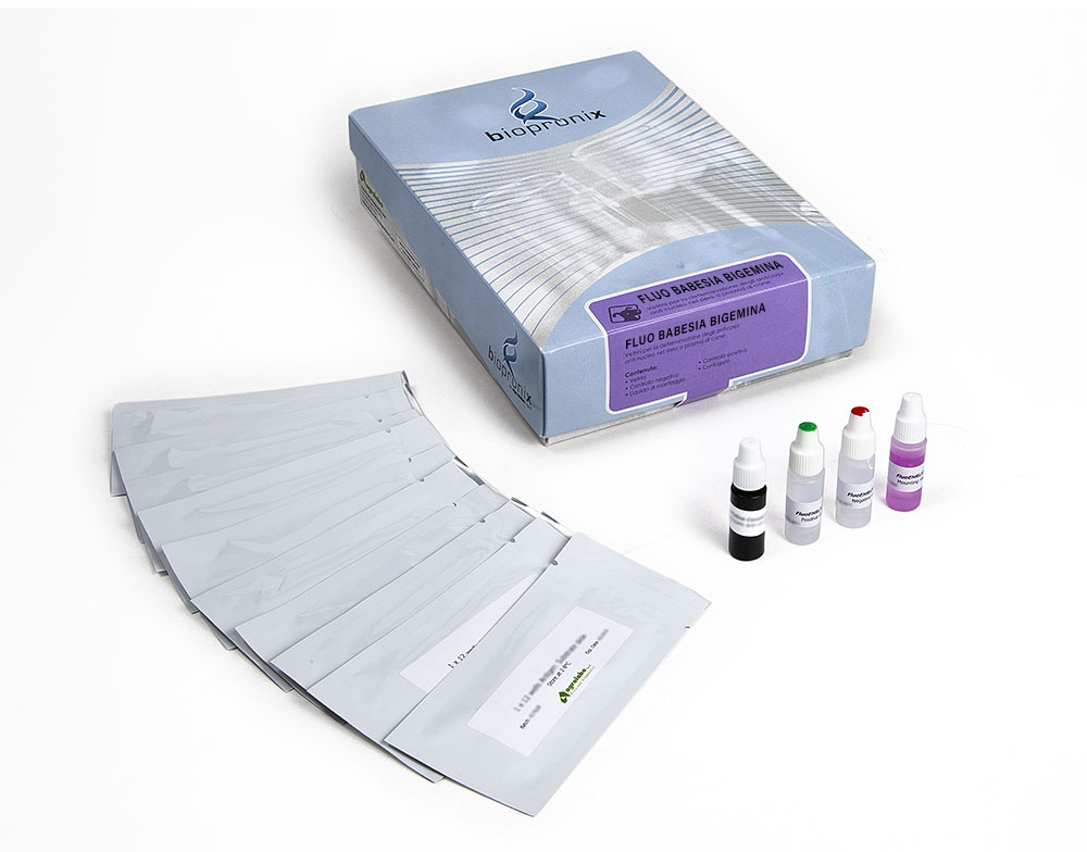
Fluo Babesia bigemina
IFA test for the detection of IgG antibodies to Babesia bigemina in bovine serum or plasma
50 slides
Complete kit
The FLUO BABESIA bigemina kit is intended for the semiquantitative detection of IgG antibodies to Babesia bigemina in bovine serum or plasma by indirect immunofluorescence assay
Bovine Babesiosis is a tick-borne protozoal disease of considerable economic importance in cattle farms in Europe. The disease is caused by Babesia bovis and Babesia bigemina, transmitted by the ticks of Boophilus spp.
Babesiosis is an infection of the erythrocytes. The genus Babesia spp. includes a group of single-celled organisms with a three-stage life cycle that includes sexual reproduction in ticks, the formation of infectious spores in the salivary gland of ticks, and asexual reproduction through bipartition in erythrocytes of vertebrate animals. Vertical transmission in ticks has been described for some of the Babesia genera. As for Babesia bovis also for Babesia bigemina the transmission in cattle occurs through the placenta.
Substrate slides consist of Teflon-masked wells containing fixed erythrocytes, which are infected with Babesia bigemina and contain the characteristic cytoplasmatic merozoites.
Bovine sera are diluted in buffered saline and incubated in the individual slide wells to allow reaction of patient antibody with Babesia antigens on the wells.
Slides are then washed to remove unreacted serum proteins, a fluorescence-labelled FITC anti-bovine IgG (conjugate) is added. This conjugate is allowed time to react with antigen-antibody complexes. The slides are washed again to remove unreacted conjugate. The resulting reactions can be visualized using standard fluorescence microscopy, where a positive reaction is seen as sharply defined apple-green fluorescent merozoites within the cytoplasm of infected erythrocytes in each well. A negative reaction is seen either as red-counterstained cells or fluorescence unlike that seen in the positive control well.
Positive reactions may then be retested at a higher dilution to determine the highest reactive (endpoint titer).
Bovine serum or plasma samples.
After collection, once coagulation has taken place, the serum is separated by centrifugation. The serum or plasma is then transferred to sterile tubes, which can be stored at + 2-8 ° C. If testing is performed after more than 5 days, samples should be frozen at -20 ° C or colder. Samples taken from acute subjects must be collected at the onset of the disease; Additional samples can be taken at intervals of two and four weeks to detect any change in antibody titer during convalescence.
The results are readable with a fluorescence microscope with FITC filter at 400X magnification.
The fluorescence pattern (shape, density, etc.) of the negative and positive control should be considered the reference model.
Reactivity patterns other than those present in the controls should be considered non-specific reactions (negative result).
The presence of anti-Babesia bigemina antibodies depends on the geographic regions and populations analysed.
