FLUO BABESIA canis
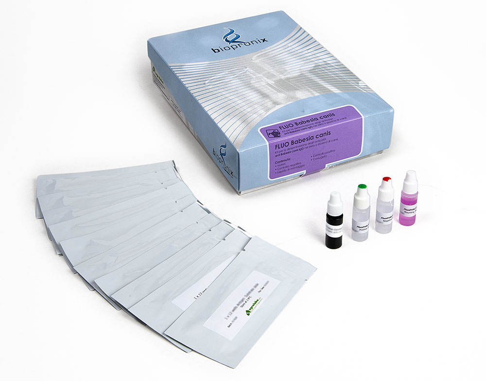
IFA kit for the detection of anti-Babesia canis IgG antibodies
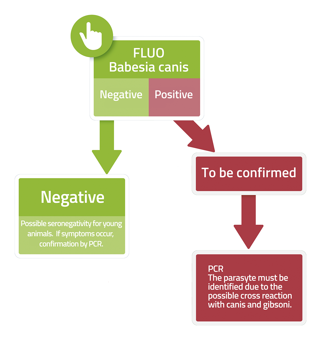
Fluo BABESIA canis is a test based on the immunofluorescence technique for the detection of IgG antibodies to Babesia canis in dog serum or plasma samples.
For many years, canine babesiosis, caused primarily by Babesia canis, was described as a predominantly hemolytic disease, characterized by parasite invasion of red blood cells, intracellular replication, and subsequent red blood cell destruction, leading to anemia.
This mechanism certainly represents a central element of pathogenesis, but recent scientific findings have demonstrated that hemolysis alone is insufficient to explain the severity of the clinical manifestations observed in patients with complicated forms.
Babesiosis is now interpreted as a systemic disease characterized by a complex interaction between the parasite, the host immune response, vascular endothelium, and the microcirculation. This model shares important similarities with some aspects of severe malaria in humans, particularly the role of endothelial damage and alterations in tissue perfusion.
1.Systemic Inflammatory Response and Multiorgan Dysfunction: SIRS and MODS
In the most severe forms of babesiosis, clinical damage is not solely due to the presence of the parasite in the red blood cells, but also to the host's inflammatory response.
- canis infection causes a strong activation of the innate immune system, involving macrophages and other immune cells. This response can lead to the increased production of pro-inflammatory mediators, including TNF-α, IL-6, and IL-1β, contributing to the development of Systemic Inflammatory Response Syndrome (SIRS). In the most severe cases, progression to Multiple Organ Dysfunction Syndrome (MODS) can result in acute renal failure, pulmonary edema, hepatic dysfunction, and neurological dysfunction, the severity of which is not always directly proportional to the level of parasitemia, as the immune response and vascular damage also significantly contribute to the progression of the disease.
2.Red Blood Cell Alterations, Cytoadhesion, and Microcirculatory Damage
One of the most interesting aspects of the pathogenesis of babesiosis concerns the changes the parasite induces on infected red blood cells.
- Red Blood Cell Membrane Modification: During its replicative cycle within the erythrocyte, Babesia alters the characteristics of the cell membrane, modifying its physical and biological properties. These transformations can facilitate the recognition and removal of infected erythrocytes by the immune system, but they can also contribute to alterations in peripheral circulation.
- Tissue Hypoxia: In some forms of severe babesiosis, interactions between infected erythrocytes, endothelial cells, and components of the coagulation system have been described. Cellular adhesion (cytoadhesion) and red blood cell aggregation can contribute to the reduction of blood flow in small vessels, leading to impaired tissue perfusion. These microcirculatory changes, combined with endothelial activation and a local inflammatory response, can lead to reduced oxygen availability to tissues (tissue hypoxia) and contribute to organ damage.
- A new perspective on the management of babesiosis: The modern interpretation of canine babesiosis goes beyond the concept of a simple parasitic anemia, recognizing the disease as a systemic immuno-inflammatory and vascular syndrome.
3.The Crosstalk Between Inflammation and Coagulation: Beyond Simple Platelet Loss
Thrombocytopenia is one of the most common hematological abnormalities in canine babesiosis, but its origin cannot be traced solely to immune-mediated platelet destruction.
Current knowledge has highlighted a central role for the interaction between the inflammatory response, vascular endothelium, and the coagulation system, a complex process that contributes to the onset of systemic complications in the most severe forms of the disease.
Endothelial Activation and Coagulation Imbalance: During Babesia canis infection, the systemic inflammatory response can lead to marked activation of the vascular endothelium. Activated endothelial cells release pro-inflammatory and pro-coagulant mediators, promoting activation of the coagulation cascade and altering the normal balance between clot formation and dissolution. This establishes a bidirectional crosstalk between the immune system and coagulation: inflammation promotes coagulation activation, while coagulation products can further amplify the inflammatory response.
- Coagulation Imbalance: In the most severe forms, the imbalance can evolve into Disseminated Intravascular Coagulation (DIC), characterized by systemic activation of coagulation with microthrombus formation, consumption of platelets and coagulation factors, and possible development of hemorrhagic disorders.
4.Proteomics and the Search for New Prognostic Biomarkers
The growing understanding of babesiosis as a systemic inflammatory and vascular disease has stimulated the search for biomarkers capable of more precisely assessing the severity of the host response and the risk of complications.
- Acute Phase Proteins (APP): C-Reactive Protein (CRP) and Serum Amyloid A (SAA) are now considered essential. They are used to assess disease severity, as they reflect the level of systemic inflammatory activation and can provide useful information in monitoring clinical progression. Persistently elevated values or a lack of reduction after initiation of antiparasitic therapy may be associated with a persistently active inflammatory response and a higher risk of complications, although they should always be interpreted in conjunction with the patient's clinical parameters.
- Apolipoprotein A1 (ApoA1): Proteomic studies have also shown that Apolipoprotein A1 (ApoA1), the main component of high-density lipoproteins (HDL), is reduced in severe forms of babesiosis, likely related to the systemic inflammatory response. This parameter is currently considered a potential negative prognostic biomarker: lower values may be associated with greater disease severity.
Bibliography
- Camacho, A.T., et al. (2005) Serum protein response and renal failure in canine Babesia annae infection. Vet Res. 36(5-6):713-22
- Jacobson, L. S., & Clark, I. A. (1994) The pathophysiology of canine babesiosis: new approaches to an old puzzle. Journal of the South African Veterinary Association, 65(3):134-45.
- Kuleš, J., et al. (2014) Identification of serum biomarkers in dogs naturally infected with Babesia canis canis using a proteomic approach. BMC Vet Res. 10:111.
- Kuleš, J., et al. (2017) Blood markers of fibrinolysis and endothelial activation in canine babesiosis. BMC Vet Res. 13(1):82.
- Matijatko, V., et al. (2007) Evidence for an acute phase response in dogs naturally infected with Babesia canis. Veterinary Parasitology, 144(3-4), 242-250.
- Schetters, T. (2019) Mechanisms involved in the persistence of Babesia canis infection in dogs. Pathogens. 8(3):94.
In dogs, babesiosis can have a hyperacute, acute, chronic or subclinical course, and there are also atypical forms. Acute disease is the most common syndrome, while hyperacute is most rare.
Hyperacute babesiosis
Typical symptoms:
- Disseminated intravascular coagulation (DIC)
- Hypothermia
- Coma
- Death
.
Acute babesiosis
Typical symptoms:
- Anorexia
- Fever
- Lethargy
- Acute haemolytic anemia
- Thrombocytopenia
- Enlargement of the spleen
- Enlargement of the lymph nodes
- Vomiting
- Jaundice
- Hematuria
.
Death can occur especially in puppies, but most animals recover from this form.
Chronic Babesiosis
There are little indications of chronic forms. The main symptoms are:
- Intermittent fever
- Decreased appetite
- Loss of weight
.
Subclinical forms are usually asymptomatic, although still a source of infection.