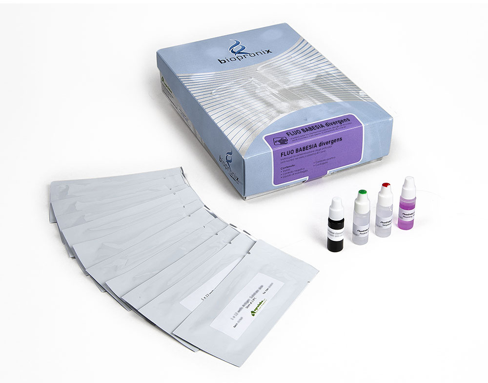
Fluo Babesia divergens
The FLUO BABESIA divergens kit can be used for the semiquantitative detection of anti-Babesia divergens IgG antibodies in bovine serum or plasma samples using an indirect immunofluorescence technique.
50 slides
Complete Kit
The FLUO BABESIA divergens kit is intended for the semiquantitative detection of IgG antibodies to Babesia divergens in bovine serum or plasma by indirect immunofluorescence assay
Babesia is a genus of pyroplasmic sporozoa belonging to the Babesiidae family, more than 100 species are known.
Babesias use ticks as a vector of transmission. They induce erythrocyte lysis therefore haemolytic anemia, the damage to the kidneys is important giving tubular necrosis, deposits and hemoglobin and edema. Symptoms include severe fever, malaise and anemia in some cases nausea, vomiting, haematuria, excessive sweating have been recorded and hepatosplenomegaly may also be present. Babesias after a first infection are neutralized by IgG antibodies until they are stored by the immune system. Numerous vaccines have been created for veterinary Babesiosis, the most effective being the one with live attenuated agents but a recombinant vaccine is being studied.
Babesia divergens infections are the most dangerous as they lead to haemoglobinuria, jaundice, pulmonary edema or even shock and kidney failure.
Substrate slides consist of Teflon-masked wells containing fixed erythrocytes, which are infected with Babesia divergens and contain the characteristic cytoplasmatic merozoites.
Bovine sera are diluted in buffered saline and incubated in the individual slide wells to allow reaction of patient antibody with Babesia antigens on the wells.
Slides are then washed to remove unreacted serum proteins, a fluorescence-labelled FITC anti-bovine IgG (conjugate) is added. This conjugate is allowed time to react with antigen-antibody complexes. The slides are washed again to remove unreacted conjugate. The resulting reactions can be visualized using standard fluorescence microscopy, where a positive reaction is seen as sharply defined apple-green fluorescent merosoit within the cytoplasm of infected erythrocytes in each field. A negative reaction is seen either as red-counterstained cells or fluorescence unlike that seen in the positive control well.
Positive reactions may then be retested at a higher dilution to determine the highest reactive (endpoint titer).
Bovine serum or plasma samples.
After collection, once coagulation has taken place, the serum is separated by centrifugation, then transferred (serum or plasma) into sterile tubes. Store at + 2-8 ° C. If testing is performed after more than 5 days, samples should be frozen at -20 ° C or colder. Samples taken from subjects with acute form must be collected at the beginning of the disease; further samples may be taken at intervals of two and four weeks to detect any change in antibody titer during convalescence.
To read the results, use the fluorescence microscope with FITC filter at 400X magnification.
The fluorescence pattern (shape, density, etc.) of the negative and positive control should be considered the reference model.
Reactivity patterns other than those present in the controls should be considered non-specific reactions (negative result).
The presence of antibodies to Babesia divergens depends on the geographic regions and populations analysed.
