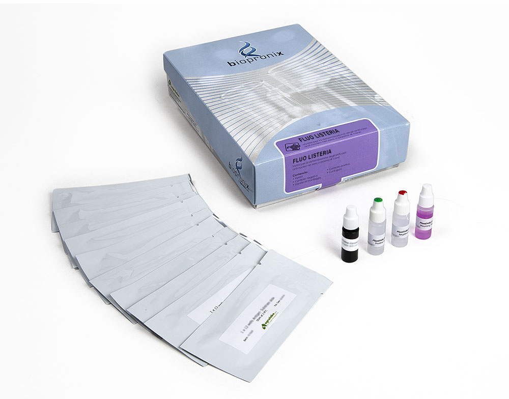
FLUO Listeria
Kit IFA for the detection of anti-Listeria monocytogenes (serotypes 1 and 4b) IgG antibodies in sheep serum or plasma
50 slides
Complete Kit
The FLUO LISTERIA monocytogenes kit is intended for the semiquantitative detection of IgG antibodies to Listeria monocytogenes (serotypes 1 and 4b) in sheep serum or plasma by indirect immunofluorescence assay
Listeriosis is a bacterial infection that can affect a large number of domestic and wild animals (mammals, birds, fish and crustaceans). The disease is spread all over the world.
Listeria monocytogenes is a Gram positive rod-shaped bacterium. It is a saprophyte, has a high tenacity (resistance capacity) against drying, light, cold and heat.
There are three main forms, in addition to the clinically inapparent one: Form that attacks the central nervous system: especially in young animals, but also in adults.
The following symptoms occur: high fever, conjunctivitis, central nervous system disorders such as opisthotonus, facial paralysis, teeth grinding, unnatural head posture, decubitus, coma. The histological picture of meningoencephalitis caused by listeriosis is characterized by perivascular infiltration of round cells in the brain stem (medulla oblongata, pons). In young animals, the infection often manifests itself as septicemia. At the end of gestation miscarriages can occur.
The intestinal tract of humans and animals represents the reservoir of the pathogen. The result is a contamination of the soil, sewage and plants. The pathogen survives for a long time in the soil. Infection occurs through the ingestion of contaminated forage (insufficiently soured ensiled). Contact or dirt infections are rare. Diaplacental transmission from the mother to the newborn animal is possible. The pathogen is secreted in milk and aborted material.
Substrate slides consist of Teflon-masked wells containing cells infected by Listeria monocytogenes (in the upper line by Listeria serotype 1 and in the lower line by Listeria serotype 4b).
Sheep sera are diluted in buffered saline and incubated in both individual slide wells to allow reaction of patient antibody with both Listeria antigens on the wells.
Slides are then washed to remove unreacted serum proteins, a fluorescence-labelled FITC anti-sheep IgG (conjugate) is added. This conjugate is allowed time to react with antigen-antibody complexes.
The slides are washed again to remove unreacted conjugate. The resulting reactions can be visualized using standard fluorescence microscopy, where a positive reaction is seen as listeria that show a clear, sharply defined apple-green fluorescence. A negative reaction is seen as a red-greyish colour of the listeria or as a fluorescence unlike that seen in the positive control well.
Positive reactions may then be retested at a higher dilution to determine the highest reactive (endpoint titer).
Sheep serum or plasma samples.
After collection, once coagulation has taken place, the serum is separated by centrifugation, then transferred to sterile tubes. Store at + 2-8 ° C. If testing is performed after more than 5 days, samples should be frozen at -20 ° C or colder. Samples taken from subjects with acute form must be collected at the beginning of the disease; further samples may be taken at intervals of two and four weeks to detect any change in antibody titer during convalescence.
To read the results, use the fluorescence microscope with FITC filter at 400X magnification.
The fluorescence pattern (shape, density, etc.) of the negative and positive control should be considered the reference model.
Reactivity patterns other than those present in the controls should be considered non-specific reactions (negative result).
The presence of antibodies to Listeria monocytogenes depends on the geographic regions and populations analysed.
